Domain             : Computer Vision, Machine Learning
Sub-Domain         : Deep Learning, Image Recognition
Techniques         : Deep Convolutional Neural Network, Transfer Learning, ImageNet, Auto ML, NASNetMobile
Application        : Image Recognition, Image Classification, Medical Imaging
1. Detected Cancer from microscopic tissue images (histopathologic) with Auto ML (Google’s “NASNet”).
2. For training, concatenated global pooling (max, average), dropout and dense layers to the output layer for final output prediction.
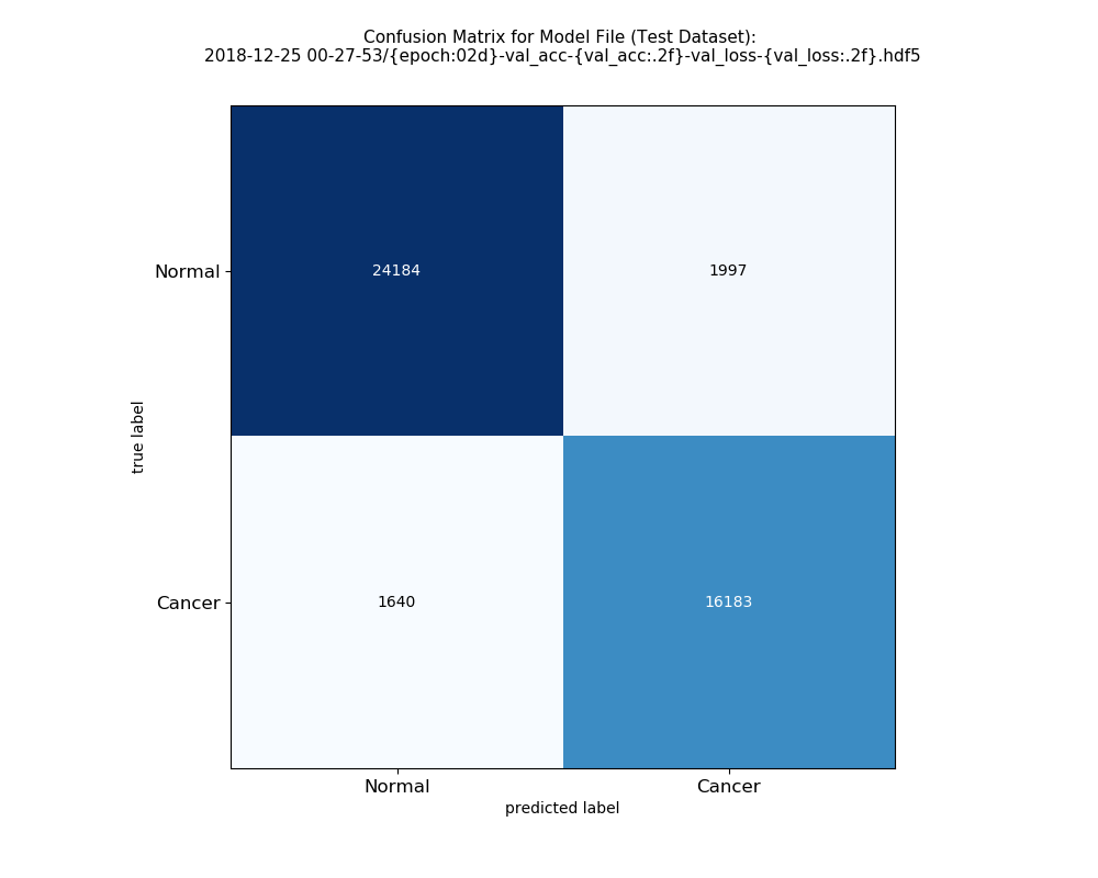
3. Attained testing accuracy of 93.72% and loss 0.30 on 250K+ (6.5GB+) image cancer dataset.
GitHub Link      : Histopathologic Cancer Detection(GitHub)
GitLab Link      : Histopathologic Cancer Detection(GitLab)
Portfolio        : Anjana Tiha's Portfolio
Dataset Name     : Histopathologic Cancer Detection
Dataset Link     : Histopathologic Cancer Detection (Kaggle)
                 : PatchCamelyon (PCam) (GitHub)
                 : CAMELYON16 challenge Dataset (Original Dataset)

Original Paper   : Diagnostic Assessment of Deep Learning Algorithms for Detection of Lymph Node Metastases in Women With Breast Cancer
                   Authors: Babak Ehteshami Bejnordi, Mitko Veta, Paul Johannes van Diest
                   JAMA (The Journal of the American Medical Association)
                   Ehteshami Bejnordi B, Veta M, Johannes van Diest P, et al. Diagnostic Assessment of Deep Learning Algorithms for Detection of Lymph Node Metastases in Women With Breast Cancer. JAMA. 2017;318(22):2199–2210. doi:10.1001/jama.2017.14585
Dataset Name            : Histopathologic Cancer Detection
Number of Class         : 2
| Dataset Subtype | Number of Image | Size of Images (GB/Gigabyte) |
|---|---|---|
| Total | 220,025 | 5.72 Gigabyte (GB) |
| Training | 132,016 | 3.43 Gigabyte (GB) |
| Validation | 44,005 | 1.14 Gigabyte (GB) |
| Testing | 44,004 | 1.14 Gigabyte (GB) |
Machine Learning Library: Keras
| Current Parameters | Value |
|---|---|
| Base Model | NashNetLarge |
| Optimizers | Adam |
| Loss Function | categorical_crossentropy |
| Learning Rate | 0.0001 |
| Batch Size | 16 |
| Number of Epochs | 2 |
| Training Time | 4.5 hour (270 min) |
| Dataset | Training | Validation | Test |
|---|---|---|---|
| Accuracy | 94.74% | 93.62% | 93.72% |
| Loss | 0.14 | 0.30 | 0.30 |
| Precision | --- | --- | 89.02% |
| Recall | --- | --- | 90.80% |
| Roc-Auc | --- | --- | 91.59% |
| Parameters (Experimented) | Value |
|---|---|
| Base Models | NashNet(NashNetLarge, NashNetMobile), InceptionV3 |
| Optimizers | Adam, SGD |
| Loss Function | categorical_crossentropy, binary_crossentropy |
| Learning Rate | 0.0001, 0.00001, 0.000001, 0.0000001 |
| Batch Size | 16, 32, 64, 128, 256 |
| Number of Epochs | 2, 4, 6, 10, 30, 50, 100 |
| Training Time | 4.5 hour (270 min), 1 day (24 hours), 2 days (24 hours) |
 See More Images

Languages               : Python
Tools/IDE               : Anaconda
Libraries               : Keras, TensorFlow, Inception, ImageNet
Duration                : November 2018 - Current
Current Version         : v1.0.0.3
Last Update             : 12.24.2018