Handheld Space Microscope
Health Medicine and Biotechnology
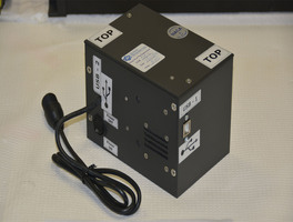
Handheld Space Microscope (MSC-TOPS-131)
Offers submicron resolution in a rugged rechargeable platform
Overview
Innovators at NASA Johnson Space Center have developed a handheld digital microscope to fill the critical microscopy needs of human space exploration by providing flight crews in situ hematological diagnostic and tracking ability to assess and monitor crew health in the absence of gravity. Although currently in use aboard the International Space Station (ISS) to work in conjunction with NASA’s handheld slide staining system, the microscope may have numerous applications here on Earth.
The microscope is entirely self-contained, and includes optics, illumination, high-resolution imaging hardware, wireless enabled single board computer with scalable power and memory, and rechargeable battery. The microscope also acts as an internet access point and connects via Bluetooth to smart devices for wireless image transfer and remote control.
The microscope is durable enough to support field use while providing submicron imaging that would typically necessitate the use of larger more expensive benchtop microscopes. Cost of manufacturing the microscope may be relatively inexpensive through the utilization of 3D-printed components, and COTS hardware such as interchangeable microscope objectives.
The handheld digital microscope is at technology readiness level (TRL) 8 (actual system completed and "flight qualified" through test and demonstration), and is now available to license. NASA does not manufacture products for commercial sale.
The Technology
The handheld digital microscope features a 3D-printed chassis to house its hardware, firmware, and rechargeable Li-ion battery with built-in power management. It incorporates an internal stainless-steel cage system to enclose and provide mechanical rigidity for the optics and imaging sensor. To reduce the microscope’s size, yet retain high spatial resolution, engineers devised an optical light path that uniquely folds back on itself using high reflectivity mirrors, thus significantly reducing internal volume.
Imaging control and acquisition is performed using a secure web-based graphical user interface accessible via any wireless enabled device. The microscope serves as its own wireless access point thus obviating the need for a pre-existing network. This web interface enables multiple simultaneous connections and facilitates data sharing with clinicians, scientists, or other personnel as needed. Acquired images can be stored locally on the microscope server or on a removable SD card. Data can be securely downloaded to other devices using a range of industry standard protocols.
Although the handheld digital microscope was originally developed for in-flight medical diagnosis in microgravity applications, prototypes were thoroughly ground-tested in a variety of environments to verify the accurate resolve of microbial samples for identification and compo-sitional analysis for terrestrial field use. Owing to its portability, other applications demanding rapid results may include research, education, veterinarian, military, contagion disaster response, telemedicine, and point-of-care medicine.

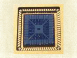
 allows quick viewing of biological cells such as this blood smear.")
Benefits
- High resolution: resolves features to just below one micron
- Portable: rechargeable microscope is palm-sized and lightweight
- Rugged construction: employs 3D-printed polycarbonate exoskeleton; utilizes stainless steel optics cage
- Remote accessible: Bluetooth/WiFi enabled to enhance data sharing for remote analysis
- Convenient data storage: features internal storage with removable SD card
- Low production cost: utilizes COTS hardware and 3D printed components
Applications
- Academic research
- Disaster areas
- Healthcare
- Military
- Natural sciences
- Other remote research and field work
Technology Details
Health Medicine and Biotechnology
MSC-TOPS-131
MSC-27481-1
MSC-27483-1
MSC-27483-2
|
Tags:
|
Similar Results

Handheld Slide Staining System for Microscopy
To stain a specimen slide, one or more liquid reagents are injected via the dispenser into the slide staining device via a syringe port. The volume of a given reagent is determined by adjustable settings on the dispenser, so that when connected to the staining device, initiates a thin film over the slide. The dispensing device uses only a fraction of the reagents typically used in non-sealed environments. Medical grade polyvinyl alcohol sponges have been incorporated into the dispenser to provide additional fluid containment and retention during the staining procedure. Furthermore, the dispenser can recall excess reagent, minimizing reagent use until refill.
The slide staining device is composed of an upper and lower section held together and aligned by use of Nd magnets. With the device open, a specimen slide is positioned upon a silicone gasket that sits within a recess in the lower section. When the device is closed, the silicone gasket in the upper section applies a seal to the slide forming a cavity that allows the slide to be exposed to reagents injected from the connected dispenser creating a stain through the use of capillary forces. Although originally designed for use in microgravity, the slide staining system also works in gravity environments.
Numerous applications may exist for this technology, particularly in hematology and cellular biology. Other applications could be considered for academic research, veterinary field use, military, disaster stricken and remote environments or where fine control of fluid delivery, removal, and management is desired.
The slide staining system is at technology readiness level (TRL) 8 (actual system completed and "flight qualified" through test and demonstration), and are now available to license. Please note that NASA does not manufacture products itself for commercial sale.
Compact Science Experiment Module
The Compact Science Experiment Module (CSEM) provides a suitable experiment platform consisting of an enclosure that contains all the required components to perform science experiments that can house either living biological samples or other samples on both the ground and on the International Space Station (ISS). The invention provides required instrumentation for video capture and data storage, environmental monitoring, inclusive of sensing temperature in degree Celsius, relative humidity as a percentage, carbon dioxide in parts per million and oxygen in percentage format. Data can be stored within the module and retrieved after the experiment or can be transmitted to the ground as for example from the ISS, by connecting to the ISS telemetry system.
The Compact Life Science Experiment Module has been fully developed at NASA Ames Research Center and tested on the ISS. In general, fruit fly studies can provide information about the effects of spaceflight at the biochemical, cellular and organismal levels. Using fruit fly spaceflight hardware, researchers are able to investigate the role of spaceflight on development, growth, reproduction, aging, neurobehavioral responses, immunity, heart function, etc. The fruit fly genome matches the human disease genome by almost 77%, and flies have, therefore, been a useful tool for scientists to understand the genetics, and molecular biology of more complex biological systems like humans. The Compact Science Experiment Module is extremely adaptable to other model specimens and samples as well, and has also flown plant experiments on the ISS. The software can be tailored to accommodate different experiment scenarios by adjusting video imaging times, LED light cycles, data storage and telemetry etc.
Portable Medical Diagnosis Instrument
The technology utilizes four cutting-edge sensor technologies to enable minimally- or non-invasive analysis of various biological samples, including saliva, breath, and blood. The combination of technologies and sample pathways have unique advantages that collectively provides a powerful analytical capability. The four key technology components include the following: (1) the carbon nanotube (CNT) array designed for the detection of volatile molecules in exhaled breath; (2) a breath condenser surface to isolate nonvolatile breath compounds in exhaled breath; (3) the miniaturized differential mobility spectrometer (DMS) -like device for the detection of volatile and non-volatile molecules in condensed breath and saliva; and (4) the miniaturized circular disk (CD)-based centrifugal microfluidics device that can detect analytes in any liquid sample as well as perform blood cell counts. As an integrated system, the device has two ports for sample entry a mouthpiece for sampling of breath and a port for CD insertion. The breath analysis pathway consists of a CNT array followed by a condenser surface separating liquid and gas phase breath. The exhaled breath condensate is then analyzed via a DMS-like device and the separated gas breath can be analyzed by both CNT sensor array again and by DMS detectors.
Micro-Organ Device
The NASA developed Micro-Organ Device (MOD) platform technology is a small, lightweight, and reproducible in vitro drug screening model that can inexpensively biomimic different mammalian tissues for a multitude of applications. The technology is automated and imposes minimal demands for resources (power, analytes, and fluids). The MOD technology uses titanium tetra(isopropoxide) to bond a microscale support to a substrate and uses biopattering and 3D tissue bioprinting on a microfluidic microchip to eliminate variations in local seeding density while minimizing selection pressure. With the MOD, pharmaceutical companies can test more candidates and concentrate on those with more promise therefore, reducing R&D overall cost.
This innovation overcomes major disadvantages of conventional in vitro and in vivo experimentation for purposes of investigating effects of medicines, toxins, and possibly other foreign substances. For example, the MOD platform technology could host life-like miniature assemblies of human cells and the effects observed in tests performed could potentially be extrapolated more readily to humans than could effects observed in conventional in vitro cell cultures, making it possible to reduce or eliminate experimentation on animals. The automated NASA developed technology with minimal footprint and power requirements, micro-volumes of fluids and waste, high throughput and parallel analyses on the same chip, will advance the research and development for new drugs and materials.

Robotic Inspection System for Fluid Infrastructures
The Robotic Inspection System improves the inspection of deep sea structures such as offshore storage cells/tanks, pipelines, and other subsea exploration applications. Generally, oil platforms are comprised of pipelines and/or subsea storage cells. These storage cells not only provide a stable base for the platform, they provide intermediate storage and separation capability for oil. Surveying these structures to examine the contents is often required when the platforms are being decommissioned. The Robotic Inspection System provides a device and method for imaging the inside of the cells, which includes hardware and software components. The device is able to move through interconnected pipes, even making 90 degree turns with minimal power. The Robotic Inspection System is able to display 3-dimentional range data from 2-dimensional information. This inspection method and device could significantly reduce the cost of decommissioning cells. The device has the capability to map interior volume, interrogate integrity of cell fill lines, display real-time video and sonar, and with future development possibly sample sediment or oil.

