The goal of cecelia is to simplify image analysis for immunologists
and integrate static and live cell imaging with flow cytometry data. The
package primarily builds upon napari and
shiny. Our aim was to combine shiny’s
graph plotting engine with napari’s image display.
This package is pre-alpha
This package currently only works on Unix systems. For Windows
system, or if you prefer a containerised version, we also have a Docker
image.
We designed cecelia to also process jobs on the HPC (High
Performance Computing) system. We currently only support Slurm as a
scheduler. If you want to set this up on your system - please open an
issue and we will get you started.
(Optional) All components can be packaged within a conda environment.
We recommend to install
miniconda if you
want to keep a separate environment. If you opt for this, then you
should also install RStudio within that conda environment.
conda create -y -n r-cecelia-env -c conda-forge python=3.9
conda activate r-cecelia-env
conda install -y -c conda-forge r-base=4.1.2 rstudio
rstudio
You can install the development version of cecelia like so:
if (!require("remotes", quietly = TRUE))
  install.packages("remotes")
remotes::install_github("schienstockd/cecelia", Ncpus = 4, repos = "https://cloud.r-project.org")
For first time users, you will need to define base directory where
configuration files, models and the shiny app will be stored. cecelia
depends on a python environment which needs to be created. There are
multiple options available depending on how you would like to use the
app:
-
imageFor image analysis on Desktop -
image-noguiFor image processing without GUI -
flowFor flow cytometry analysis
library(cecelia)

# install App requirements
# (i) they are not needed when using only markdown files or on HPC
cciaAppRequirements(repos = "https://cloud.r-project.org")

# install Bioconductor requirements
cciaBiocRequirements(repos = "https://cloud.r-project.org")

# setup cecelia directory
cciaSetup("~/path/to/cecelia")

# create conda environment
cciaCondaCreate()
# cciaCondaCreate(envType = "image-nogui") # to use without gui
# cciaCondaCreate(envType = "flow") # for flow based only

# download models for deep-learning segmentation
cciaModels()

# create app
cciaCreateApp()
You have to adjust the parameters in ~/path/to/cecelia/custom.yml to
your system. You need download/install:
-
ImageJif using Spot segmentation
For ImageJ, activate the following update sites:
-
IJPB-plugins
-
3D-ImageJ-Suite
-
Bio-Formats
default:
  dirs:
    bftools: "/Applications/BFTools"
    bioformats2raw: "/Applications/glencoe/bioformats2raw-0.3.0"
    projects: "/your/project/directory/"
  volumes:
    SSD: "/your/ssd/directory/"
    home: "~/"
    computer: "/"
  python:
    conda:
      env: "r-cecelia-env"
      source:
        env: "r-cecelia-env"
  imagej:
path: "/Applications/Fiji.app/Contents/MacOS/ImageJ-macosx"library(cecelia)
cciaUse("~/path/to/cecelia", initJupyter = TRUE)
cciaRunApp(port = 6860)-
Create a Project for
staticorliveanalysis. -
Images have to be imported as
OME-ZARR. ChooseImport Imagesand create anExperimental Set. It is helpful if all images within this set have the same colour combinations. Add Images. Select all images you want to import and chooseOME-ZARR. Select the requiredpyramid scalesandrunthe task. -
Select
Image Metadataand clickLoad Metadatato load the channel information. You can assign channels either one by one by selecting a channel andSpecify Value>Assign Value. Alternatively, you can give a list of channels in the box and clickAssing channels. You can add further experimental attributes byCreate Attributeand adding respecive values for the individual images. -
Select
Cell Segmentationto segment your images. -
Select
Population Gatingto use flow cytometry like gating to define populations. SelectPopulations Clusteringto use cluster algorithms to define populations. -
Select
Spatial Analysisto define spatial neighbourhoods, detect clustering cells or detect contact between cells.
library(cecelia)
cciaUse("~/path/to/cecelia", initJupyter = FALSE)
cciaRunApp(port = 6860)-
Create a Project for
flowanalysis. -
FCSfiles can be imported either from raw or other sources such asFlowJo. They will converted into anAnndatato perform clustering and aGatingSetto perform manual gating.
Compensation controls can be added into one experimental set which can
be used by autospill to calculate a compensation matrix. This matrix
can then be applied to the other samples.
- The rest of the pipeline for
gatingandplottingis the same as for image analysis.
All Processing available in the app can be done from RMarkdown as
well. Every image is ReactivePersistentObject whose state is saved in
an RDS file and is reactive in a shiny app context. These objects
can be loaded and manipulated as such:
library(cecelia)
cciaUse("~/path/to/cecelia")

# set test variables
pID <- "pEdOoZ"   # project ID
versionID <- 2    # version ID
uID <- "tPl6da"   # image ID

# init ccia object
cciaObj <- initCciaObject(
  pID = pID, uID = uID, versionID = versionID, initReactivity = FALSE
)

funParams <- list(
  pyramidScale = 4,
  dimOrder = "",
  createMIP = FALSE,
  rescaleImage = FALSE
)

cciaObj$runTask(
  funName = "importImages.omezarr",
  funParams = funParams,
  runInplace = TRUE
)- Create project
- Import image
- Assign channel names
- Segment cells
With conventional confocal microscopy it is not always possible to
include a nuclear stain for cell segmentation. In this case, we can
check whether cellpose or a sequence of morphological filters which
segments donut- and blob-like objects (donblo) works for a partiular
image.
cellposeis a good choice for most cases. We usecellposeto segment a single merged image. We can create a sequence of merged images to create individual segmentations if necessary.
- In this case,
cellposedid not capture some of the more dense and noisy cells. We have implemented a simple sequence of morphological filters with subsequent spot detection and segmentation inImageJusingTrackMateand the3D Image Suite. The quality of the segmentation is lower thancellposebut it will capture more cells, such as theyellow XCR1+ DCswithin theT cell zone.
- Gate cells
Cell populations can be created using clustering or gating. Gating
cell populations will give you more control when using fewer markers.
Clustering will be more beneficial when using multiplex images to
identify multiparameter cell populations. In this case, we will utilise
gating. We have to create a GatingSet from the label properties.
After this we can open the image and do sequential gating for T cells,
Macrophages, cDC1 and cDC2.
- Create spatial neighbours
We can use these populations to create spatial neighbours.
- Custom plotting of interactions
Our aim is to provide custom plots within cecelia but this is still in
development. The following is illustrating how to use the generated
populations for customised plotting within RMarkdown. This simple
example shows the interactions between T cells and cDC1 in the
T cell zone.
library(ggplot2)
library(tidyverse)

library(cecelia)
cciaUse("~/path/to/cecelia")

# set test variables
pID <- "s3n6dR"   # project ID
versionID <- 1    # version ID
uID <- "LvfcHB"   # image ID

# init ccia object
cciaObj <- initCciaObject(
  pID = pID, uID = uID, versionID = versionID, initReactivity = FALSE
)

# get populations
popDT <- cciaObj$popDT("flow", pops = cciaObj$popPaths("flow"))

# get spatial information
spatialDT <- cciaObj$spatialDT()

# join pops
spatialDT[popDT[, c("label", "pop")],
          on = c("to" = "label"),
          pop.to := pop]
spatialDT[popDT[, c("label", "pop")],
          on = c("from" = "label"),
          pop.from := pop]

# filter same type associations
spatialDT <- spatialDT[pop.to != pop.from]

# get interaction frequencies
freqRegions <- spatialDT %>%
  group_by(pop.from, pop.to) %>%
  summarise(n = n()) %>%
  mutate(freq = n/sum(n)) %>%
  drop_na() %>%
  ungroup() %>%
  complete(pop.from, pop.to, fill = list(freq = 0))

ggplot(freqRegions,
       aes(pop.from, pop.to)) +
  theme_classic() +
  geom_tile(aes(fill = freq), colour = "white", size = 0.5) +
  viridis::scale_fill_viridis(
    breaks = c(0, 0.5),
    labels = c(0, 0.5)
  ) +
  theme(
    legend.title = element_blank(),
    legend.key.size = unit(3, "mm"),
    axis.text.x = element_text(angle = 45, hjust = 1, vjust = 1)
) + xlab("") + ylab("")- Population detection by clustering
We have incorporated UMAP and Leiden to detect populations by
clustering. Clustering can be done on individual images or combined on
multiple images from the same set. If multiple images are used, the
clustering results will be written back to the original individual
images and subsequent analysis such as neighbour detection can be done
on the individual images again. This is useful when processing a
batch of images that have the same staining.
It is also possible to do sequential clustering, as a kind of
multidimensional gate, by selecting Root populations from which to
calculate clusters. When you do not tick Keep other populations the
other populations will be removed during clustering. So if you want to
sequential clusters, please tick that box.
- Create project
- Import image
- Assign channel names
- Segment cells
We commonly use fluorescently stained cells for two-photon and
histology imaging. We trained a cellpose model, called
ccia Fluorescent, to detect these fluorescent cells as the pre-trained
models could not segment them. We can use this model to sequentially
segment dendritic cells (stained with TRITC) and T cells.
Sometimes the segmentation can benefit from a small morphological
filter. In this case we can use a Gaussian filter of 1 to improve
segmentation results.
- Gate cells
During imaging of thick tissue slices that were stained with antibodies
it can happen that staining intensity varies across the depth of the
tissue due to antibody penetration or differences in light properties
and scattering within the tissue or increase of laser power or gain due
to reduction of signal with increasing depth. For this reason, we
incorporated depth correction by fitting polynomial function to the
signal across depth.
We can use gating to identify TRITC+ dendritic cells and T cells.
In this image, we can also gate on CD68+ T cells.
- Detect spatial interactions
For 3D objects it is helpful to generate 3D meshes during
segmentation. These meshes can then be utilised to detect clusters of
cells and interactions between cells. Then we can check whether
CD69+ T cells are in contact with migrating TRITC+ cells and
visualise these in napari.
library(ggplot2)
library(tidyverse)

library(cecelia)
cciaUse("~/path/to/cecelia")

# set test variables
pID <- "4UryU2"   # project ID
versionID <- 1    # version ID
uID <- "jpVjeh"   # image ID

# init ccia object
cciaObj <- initCciaObject(
  pID = pID, uID = uID, versionID = versionID, initReactivity = FALSE
)

# get populations
popDT <- cciaObj$popDT(
  "flow", pops = c("/nonDebris/gBT/clustered", "/nonDebris/gBT/non.clustered"),
  includeFiltered = TRUE)

# get summary
summaryToPlot <- popDT %>%
  group_by(pop, `flow.cell.contact#flow./nonDebris/others/TRITC`) %>%
  summarise(n = n()) %>%
  mutate(
    freq = n/sum(n),
    pop = str_extract(pop, "[^\\/]+$")
    )

# plot frequencies
ggplot(summaryToPlot) +
  aes(1, freq, fill = `flow.cell.contact#flow./nonDebris/others/TRITC`) +
  theme_classic() +
  geom_bar(stat = "identity", width = 1) +
  coord_polar("y", start = 0) +
  xlim(0, 1.8) +
  facet_wrap(.~pop, nrow = 1) +
  ggtitle("TRITC contact") +
  theme(
    axis.text = element_text(size = 5),
    axis.text.x = element_blank(),
    axis.ticks.x = element_blank(),
    axis.text.y = element_blank(),
    axis.ticks.y = element_blank(),
    legend.title = element_blank(),
    legend.position = "bottom",
    axis.title.x = element_blank(),
    axis.title.y = element_blank()
)- Create project
- Import image
- Assign channel names
- Autofluorescence and drift correction
We correct autofluorescence by dividing channels from each other. In
this example there are only two which we can use for channel correction.
The same module function will also do drift correction based on
phase cross correlation.
We will use the AF generated channel that is generated during
autofluorescence correction.
[IMAGES HERE]
- Segment cells
We can utilise cellpose and the ccia Fluorescent model to segment
both cell types.
- Track cells
We incorporated
btrack
to track segmented cells. Filters can be used to filter based on
object measurements.
- Extract cell behaviour
We utilised HMM (Hidden markov model) to extract behaviour from the
generated tracks. This analysis extracts a defined number of cellular
behaviours based on shape and movement parameters. For this method,
we combine tracking data from multiple images - therefore, we need to
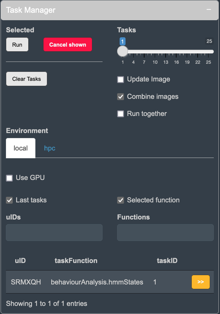
tick the box to Combine images when running the task. This will run
the task with the selected images from the set.
We can visualise these behaviours on the image by creating a
filtered population.
Then we can map these behaviours in time and space.
library(ggplot2)
library(tidyverse)

library(cecelia)
cciaUse("~/path/to/cecelia")

# set test variables
pID <- "kicbHw"   # project ID
versionID <- 1    # version ID
uID <- "SRMXQH"   # image ID

# init ccia object
cciaObj <- initCciaObject(
  pID = pID, uID = uID, versionID = versionID, initReactivity = FALSE
)

# get popDTs for set
popDTs <- cciaObj$popDT(
  popType = "live", pops = c("cellA/tracked", "cellB/tracked"),
  includeFiltered = TRUE, flushCache = TRUE)

# get frequencies of HMM at time points
hmmTime <- popDTs %>%
  dplyr::filter(
    !is.na(live.cell.hmm.state.default),
    live.cell.track.clusters.default != "NA"
    ) %>%
  group_by(uID,
    centroid_t,
    live.cell.hmm.state.default
    ) %>%
  summarise(n = n()) %>%
  mutate(
    live.cell.hmm.state.default = as.factor(live.cell.hmm.state.default),
    freq = n/sum(n)
    )

time.interval <- cciaObj$cciaObjects()[[1]]$omeXMLTimelapseInfo()$interval

ggplot(hmmTime,
       aes((centroid_t * time.interval), freq,
           color = live.cell.hmm.state.default,
           fill = live.cell.hmm.state.default,
           )) +
  geom_smooth() +
  theme_classic() +
  scale_color_brewer(name = NULL, palette = "Dark2") +
  scale_fill_brewer(name = NULL, palette = "Dark2") +
  theme(
    legend.title = element_blank(),
    legend.position = "bottom"
    ) +
  xlab("Time (min)") + ylab("HMM frequency") +
  ylim(0, 1) + facet_grid(uID~.)

# get frequencies of hmm states at time points
hmmSpace <- copy(popDTs) %>%
  dplyr::filter(!is.na(live.cell.hmm.state.default))

# get density colours
hmmSpace$density <- ""
for (i in unique(hmmSpace$uID)) {
  for (j in unique(hmmSpace$live.cell.hmm.state.default)) {
    x <- hmmSpace[hmmSpace$uID == i & hmmSpace$live.cell.hmm.state.default == j,]

    hmmSpace[hmmSpace$uID == i & hmmSpace$live.cell.hmm.state.default == j,]$density <- .flowColours(
      x$centroid_x, x$centroid_y)
  }
}

ggplot(hmmSpace, aes(centroid_x, centroid_y)) +
  theme_classic() +
  plotThemeDark(
    fontSize = 8,
    legend.justification = "centre"
    ) +
  geom_point(
    color = hmmSpace$density, size = 0.5
    ) +
  scale_color_brewer(name = NULL, palette = "Set3") +
  coord_fixed() +
  theme(
    axis.text.x = element_blank(),
    axis.ticks.x = element_blank(),
    axis.text.y = element_blank(),
    axis.ticks.y = element_blank(),
    axis.title.x = element_blank(),
    axis.title.y = element_blank(),
    legend.title = element_blank(),
    legend.position = 'bottom'
    ) +
  facet_grid(uID~live.cell.hmm.state.default)


